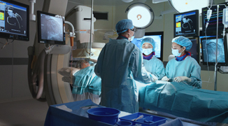
The Cardiovascular Suite is a fully integrated, purpose-built interventional facility equipped with current-generation angiography technology and imaging systems. Unlike a standard operating theatre or a basic catheterisation laboratory, it is designed as a hybrid environment — one capable of supporting both diagnostic imaging procedures and surgical interventions within the same controlled space.
Many cardiovascular procedures begin as diagnostic — a coronary angiogram to identify a blockage, for instance — and may proceed directly to intervention in the same setting if findings warrant it. Having full surgical and imaging capabilities co-located eliminates the need to transfer a patient mid-procedure, which reduces risk, shortens procedure time, and allows the clinical team to respond immediately to intraoperative findings.
The suite is staffed by a team of specialist cardiologists, interventional radiologists, vascular surgeons, cardiac nurses, and radiographers — each working within a system that is built to support complex, high-precision procedures from start to monitored recovery.
High-definition fluoroscopic imaging for real-time visualisation of blood vessels, coronary arteries, and cardiac structures during diagnostic and interventional procedures. Supports guidewire and catheter navigation with precision imaging.
Three-dimensional rotational imaging integrated within the suite, allowing cross-sectional volumetric views of vascular and cardiac anatomy without transferring the patient to a separate CT scanner. Enables pre-procedural planning and intraoperative confirmation.
A full surgical-grade operating environment — sterile field, anaesthetic support, specialist lighting, and resuscitation equipment — co-located with the angiography system, allowing seamless transition from image-guided intervention to open surgical access if required.
Continuous intraoperative monitoring of heart rate, blood pressure, oxygen saturation, and cardiac electrical activity throughout all procedures, displayed to the clinical team in real time on integrated monitoring systems.
The most common type of heart disease, CAD occurs when the coronary arteries that supply blood to the heart become narrowed or blocked due to plaque buildup. It can lead to chest pain (angina), heart attacks, and heart failure.
These are irregular heart rhythms caused by problems with the heart’s electrical system. Common types include atrial fibrillation, bradycardia, and tachycardia. Some arrhythmias can increase the risk of stroke or sudden cardiac arrest.
This occurs when one or more of the heart’s valves don’t function properly, leading to issues like stenosis (narrowing), regurgitation (leaking), or prolapse. It can affect blood flow and heart efficiency.
A condition where narrowed arteries reduce blood flow to the limbs, usually the legs. PAD increases the risk of heart attack and stroke and is often linked to atherosclerosis.
Conditions affecting the aorta, the main artery from the heart, such as an aortic aneurysm (a bulge in the artery wall), which can be life-threatening if it ruptures.
A group of conditions that affect blood flow to the brain, most commonly leading to strokes, transient ischemic attacks (TIAs), or brain aneurysms.
The defining feature of the Cardiovascular Suite is its hybrid configuration — a surgical setup and a full angiography imaging system in a single integrated room. This is clinically significant in ways that a standard catheterisation laboratory or a conventional operating theatre is not.
Many cardiovascular conditions benefit from procedures that span both interventional and surgical techniques. Endovascular aortic repair, for example, requires real-time fluoroscopic guidance for device deployment alongside the sterile field, surgical access capability, and anaesthetic support that an operating theatre provides. Transcatheter-based structural heart procedures require high-definition imaging combined with surgical readiness for conversion if complications arise.
Performing these procedures in two separate rooms — first a cath lab for the imaging phase, then an operating theatre for surgical access — requires patient transfer, a change of environment, a reconfigured team, and wasted time. The hybrid suite eliminates these transitions. The clinical team, the imaging system, the surgical equipment, and the patient remain in the same space from procedure start to close.
In cardiovascular care, the ability to convert from an image-guided intervention to open surgical access — in the same room, with the same team, without delay — is not a contingency. For some procedures, it is the standard of care.
The cone beam CT capability within the suite adds a further dimension. Where conventional angiography provides two-dimensional fluoroscopic images, CBCT provides three-dimensional cross-sectional views that can be acquired on the procedure table without the patient leaving the room. This is particularly valuable for planning complex aortic and peripheral vascular interventions, confirming device position after deployment, and identifying complications that two-dimensional imaging alone might miss.
Immediately following a cardiovascular procedure, the period of highest physiological variability begins. Blood pressure stabilises, the access site is monitored for haematoma or bleeding, sedation wears off, and the patient's cardiac rhythm is observed for changes that may follow the intervention. This recovery window requires attentive, continuous monitoring — not a standard ward environment.
The Cardiovascular Suite includes a dedicated Close Monitoring Unit, functioning as a day ward adjacent to the procedure room, specifically designed for this post-procedural recovery phase. It is staffed by trained cardiac nurses who monitor vital signs, access sites, and clinical status continuously during the recovery period, with the ability to escalate immediately to the clinical team if needed.
For patients undergoing same-day procedures, the Close Monitoring Unit is typically where discharge criteria are assessed before the patient is cleared to return home. For those admitted as inpatients, it provides the bridge between the procedure room and their inpatient suite on levels 7–10.
The unit's positioning directly adjacent to the Cardiovascular Suite is deliberate. In the event that a patient's condition changes during recovery and urgent re-intervention is required, the path back to the procedure room is immediate — with no transfer through corridors or between floors.
Cardiovascular procedures range from a 20-minute diagnostic angiogram to a multi-hour interventional or hybrid surgical procedure. The journey through the suite follows a consistent structure regardless of procedure complexity.
| Before Admission | Your cardiologist or vascular surgeon will have reviewed your imaging and clinical history. Pre-procedure instructions — fasting, medication adjustments, and access site preparation — will be provided in advance. For elective procedures, pre-admission assessment confirms fitness and ensures all equipment and team members are prepared for your specific case. |
| Arrival at Level 5 | You will be received by the Cardiovascular Suite nursing team, changed into a procedure gown, and prepared for the access site (typically wrist or groin). Intravenous access is established, and baseline vital signs are documented. Your clinical team will confirm the procedure plan and answer any final questions before you enter the suite. |
| In the Suite | The procedure is performed by your specialist with the support of a scrub nurse, radiographer, and monitoring nurse. You will be awake or lightly sedated for most catheter-based procedures; general anaesthesia is used where indicated. The team communicates with you throughout and will explain what they are doing at each stage. |
| In the Close Monitoring Unit | Following the procedure, you move to the Close Monitoring Unit where nursing staff monitor your vital signs, access site, and comfort. The duration of monitoring depends on the procedure — from one to two hours for a diagnostic angiogram to several hours for more complex interventions. |
| Discharge or Inpatient Transfer | Once discharge criteria are met — stable vital signs, haemostasis at the access site, acceptable pain levels, and clinical clearance from your doctor — you will either be discharged home with written instructions or transferred to your inpatient suite if you are admitted for an overnight stay. |
| Follow-up | Your specialist will schedule a follow-up appointment to review any pathology or device check results, discuss findings, and plan next steps in your care. |
Contact: (65) 6705 2835
Location: Level 5
Operating Hours:
Mon - Fri: 8 a.m. – 5 p.m.
Sat: 8 a.m. – 1 p.m.
Sun & PH: Closed
