
MRI Dementia Biomarker Screen

What is an MRI Dementia Biomarker Screen?

Magnetic Resonance Imaging (MRI) is a painless, non-invasive imaging procedure that uses a strong magnetic field and radio waves, instead of X-rays, to create detailed pictures of the inside of the body. It is especially good at showing soft tissues such as the brain.

In people with dementia and related brain diseases, parts of the brain gradually shrink, and this often begins years before symptoms are noticed. These measurable changes in the brain are called biomarkers. A technique called brain volumetry uses the MRI images to measure the volume of different parts of the brain, such as the hippocampus, a structure that is important for memory and that is often one of the first areas to be affected in Alzheimer’s disease. The measurements are done with the help of artificial intelligence (AI) software, which maps the different structures of the brain.

Your measurements are then compared with those of people of the same age and sex who have healthy brains, and are usually shown on a percentile chart. This helps your doctor to see whether a part of your brain is smaller than expected, and by how much.
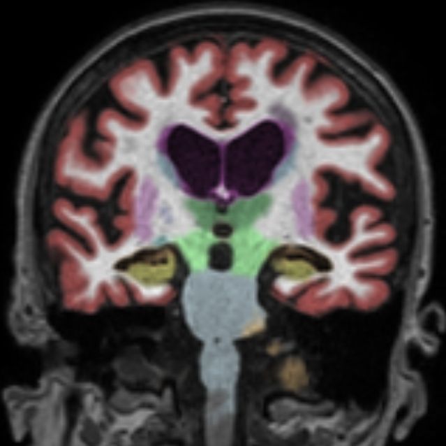


An MRI Brain with Volumetry showing different structures that have been mapped with AI.


What is the role of MRI in Dementia?

MRI is the most comprehensive imaging tool in the first assessment of a person who has symptoms that may be caused by dementia, such as memory loss. As well as looking for signs of shrinkage, it helps to find or exclude other causes of memory and thinking problems that may be treatable, such as tumors, strokes, bleeding, fluid build-up in the brain and abnormal blood vessels.
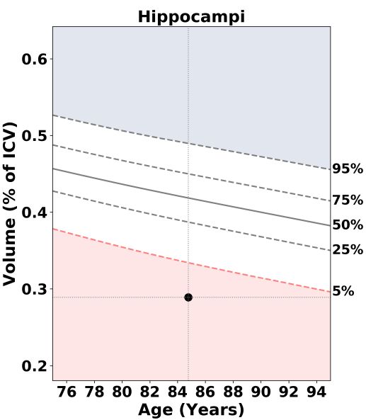


Hippocampus volume (biomarker) compared on a percentile chart.


How does it differ from a routine MRI scan?

A routine MRI scan of the brain takes pictures of the brain. An MRI brain volumetry scan is an enhanced test that takes pictures and also measures the volume of different parts of the brain. Volumetry shows shrinkage that is not easily seen on a routine MRI scan, and gives your doctor numbers to compare against what is expected for your age. It can also be repeated over time, to see whether the shrinkage is changing.

The experience for you is the same as for a routine MRI. The difference is the extra information on disease biomarkers in your report.

The scan can also show damage to the small blood vessels of the brain and narrowing of the large vessels, which is useful in the assessment of vascular dementia, a type of dementia caused by poor blood supply to the brain. An MRI scan shows the structure of the brain, while PET scans such as the F18 FDG brain scan show how active the brain is, and an amyloid PET scan looks for a specific protein linked to Alzheimer’s disease.

You may learn more about this scan by reading our aticle, Seeing What We Don't See: Dementia A-Eye.


How is the procedure performed?

Three types of dementia biomarker screening MRI are available:

  • MRI Dementia Screen: Includes an MRI brain scan and MRI volumetry. This is suitable for most people who have memory or thinking problems, or who wish to have the brain checked.
  • MRI Dementia Screen Plus: Includes an MRI brain scan, an MR angiogram (MRA) of the brain, an MRA of the carotid arteries in the neck, and MRI volumetry. The MRA scans show the blood vessels that supply the brain, and are useful for people with risk factors for vascular dementia and stroke, such as high blood pressure, diabetes, high cholesterol or smoking.
  • MRI Volumetry Only: For patients who have had a recent MRI brain scan, or for follow-up volumetry, to compare against earlier results.

Why You Might Need an MRI Dementia Biomarker Screen?

Your doctor may recommend this scan if you or a family member have:

  • Memory loss, such as forgetting recent conversations, repeating questions or misplacing things often, that affects daily life
  • Difficulty with thinking, planning, solving problems, finding words or following conversations
  • Getting lost in familiar places, or confusion about time or place
  • Changes in mood, behaviour or personality, such as becoming withdrawn, irritable, suspicious or anxious, that are out of character
  • A family history of dementia, and concern about your own memory
  • Risk factors that can affect the brain’s blood vessels, such as high blood pressure, diabetes, high cholesterol, heart disease or a previous stroke
  • A diagnosis of mild cognitive impairment or dementia, where your doctor wishes to find the cause or to follow how the brain is changing


Preparing For Your MRI Dementia Biomarker Screen

There is little preparation needed for this scan. Our team will give you specific instructions when you make your appointment. In general:

  • Eating, drinking and medicines: Unless you are told otherwise, you may eat, drink and take your medicines as usual on the day of the scan. If you will be sedated, our team will tell you whether you need to fast.
  • Remove all metal: The MRI scanner has a very strong magnet, which can pull on metal objects, damage them and cause injury to you or our staff. You will be asked to remove all metal items, such as piercings (ear, nose and body rings), jewellery such as earrings, necklaces and bracelets, watches, dentures, hearing aids, glasses, hairpins and clips, belts, coins and keys. Please also remove headscarf (tudung) pins and any wig, as some wigs contain traces of metal. Leave valuables at home, and a locker will be provided for your belongings.
  • Clothing: You will be asked to change into a hospital gown. Wear comfortable clothing without metal fasteners or zips, if possible.
  • Medical history and implants: Our staff will ask you about your health and medical history, to make sure that the scan is safe and comfortable for you. Tell our team if you have a pacemaker, defibrillator, cochlear (ear) implant, brain aneurysm clip, nerve stimulator, insulin pump, joint replacement, metal plates, screws or stents, or metal fragments in your body, for example from an injury or from work such as welding. Some implants are safe to scan, but others are not, so please bring your implant card, if you have one.
  • Tattoos and patches: Tell our staff about any tattoos, permanent make-up, magnetic products, and medicated or adhesive patches, as some contain metal.
  • Pregnancy: Tell our staff if you are pregnant or think you may be pregnant.
  • Fear of enclosed spaces: If you are afraid of enclosed spaces, inform your doctor or our team before the scan. Sedation may be arranged for you, and if so you will need an adult to take you home.
  • Bring a companion: If you or your family member has memory problems, it helps to have a family member or caregiver with you, to provide the medical history and to help you through the visit.
  • What to bring: Your doctor’s referral letter, your NRIC (or passport if you are an international patient), previous brain scan reports and images, and a list of your medications.


What Happens During an MRI Dementia Biomarker Screen?

  • The scan is performed by a radiographer who is trained in MRI, in our Diagnostic Imaging Suite, which offers a wide range of diagnostic imaging services.
  • You will be asked to change into a gown, remove all metal items and store your belongings in a locker. The radiographer will check again that you have no metal on you, and will explain the procedure.
  • You will lie flat on a motorised bed, which will move you into the scanner, head first. The scanner is a wide, open-ended tunnel. A frame (head coil) will be placed over your head, to help to receive the signals and to keep your head still.
  • You will be given earplugs or headphones, because the scanner makes loud, repetitive knocking and buzzing noises. You will be able to speak to the radiographer through an intercom, and you will be given a call button to hold.
  • It is important to keep as still as possible during the scan, because movement can blur the images. You may be asked to hold your breath briefly during some scans, such as those of the neck.
  • No contrast dye injection and no radiation are involved. If an MRA is included, it is done using the scanner alone.


What Happens After an MRI Dementia Biomarker Screen?

  • You can usually leave the hospital immediately after the scan, and return to your normal activities, including eating, drinking and taking your medicines.
  • If you were given sedation, you will rest in a recovery room until you are awake and feel well enough to go home. You should not drive, and you should have an adult accompany you home and stay with you for the rest of the day.
  • The volumetry measurements are processed by special software, and the images are reviewed and reported on by a specialist radiologist, who sends the report to your doctor. Our team will tell you when to expect your results.
  • You will be given a follow-up appointment, and your doctor will go through the results with you. If you have memory problems, it helps to have a family member or caregiver with you.


Understanding Your Results

A specialist radiologist examines your images and measures the volume of different parts of the brain, and compares them with the values expected for people of your age and sex. Your report may include the following:

  • Brain volumes: Volumes of areas such as the hippocampus and other regions are shown on a percentile chart. A low percentile means that the area is smaller than in most people of the same age and sex. Some shrinkage is a normal part of ageing, so your doctor will look at the pattern as well as the numbers.
  • Pattern of shrinkage: Different diseases tend to affect different parts of the brain. For example, shrinkage of the hippocampus and nearby areas is typical in Alzheimer’s disease, while shrinkage at the front of the brain may point to frontotemporal dementia. The pattern can help your doctor to narrow down the cause.
  • Blood vessel changes: Signs of damage to the small blood vessels, such as changes in the white matter of the brain, small old strokes or bleeds, and narrowing of the arteries seen on the MRA, suggest vascular disease, which can cause or add to memory problems, and is treated by controlling the risk factors.
  • Other findings: The scan can show other causes of memory and thinking problems that may be treated, such as tumours, strokes, fluid build-up in the brain (hydrocephalus), blood collections after head injury and abnormal blood vessels. It may also show unrelated findings that need to be followed up.
  • A normal result: This is reassuring, but it does not completely rule out early disease, because the brain may not have shrunk yet, and your doctor may recommend other tests or a repeat scan later.


Looking After Your Brain Health

  • Keep your blood pressure, blood sugar and cholesterol under control, with regular check-ups, and take your medicines as prescribed
  • Be physically active: aim for at least 150 minutes of moderate physical activity every week, as advised by your doctor
  • Keep your mind active, with reading, learning new skills, hobbies and puzzles
  • Stay socially connected with family, friends and community activities
  • Do not smoke, limit alcohol, and eat a balanced diet that is low in salt and rich in vegetables, fruit, wholegrains and fish
  • Have your hearing and eyesight checked, and use hearing aids or glasses if you need them
  • Get enough sleep, and seek help if you feel low, as depression can affect memory
  • Protect your head from injury, and prevent falls at home
  • See a doctor early if you or your family notice changes in memory, thinking or behaviour, rather than assuming that they are a normal part of ageing

You may check with your doctor or reach out to us at +65 6363 1818 to find out more. You may also send an enquiry here.

Should you feel unwell after the procedure, contact your doctor immediately or call our 24-Hour Medical Urgency Clinic