
People with memory loss or suspected dementia often undergo an MRI to exclude underlying lesions such as tumors and vascular malformations. The results of these MRIs are usually unremarkable. However, we can now obtain imaging biomarkers using visual rating and artificial intelligence (AI) to measure regional brain volumes implicated in dementia.
The earliest presenting symptom of a person suffering dementia is often memory loss. However, before memory loss, changes in the brain have already begun. These changes usually start with the deposition of abnormal proteins in the brain, followed by reduced brain metabolism and finally atrophy of the brain due to tissue destruction. Thus, brain atrophy should be present when patients present with memory loss if the cause is due to dementia. In 2011, the National Institute of Aging and the Alzheimer’s Association updated the guidelines for the diagnosis of dementia due to Alzheimer’s Disease. They added imaging biomarkers to the guidelines, which included hippocampus volume and rate of brain atrophy. Thus, the ability to detect atrophy in these structures in a person with memory loss can help in the early detection of dementia.
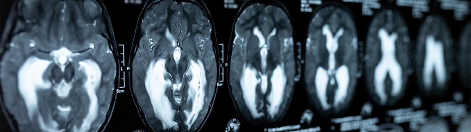
Brain atrophy’s visual rating assessment can be performed using the Global Cortical Atrophy Scale (GCA), Medial Temporal Lobe Atrophy Scale (MTA), and the Koedam Posterior Atrophy Score. An additional visual rating that is usually assessed is the Fazekas white matter lesion score that stratifies the degree of small vessel disease. Any abnormal score usually suggests that there may be some form of underlying dementia or neurodegenerative disease.
However, visual rating is limited because it relies on qualitative information, and there can be interobserver variability. Thus, it is unable to detect early changes or assess for subtle changes over time. The documentation of the results is also in broad categories that do not provide meaningful temporal comparisons or comparisons with healthy individuals.
Measurement of brain volumes (brain volumetry) using AI allows us to resolve the limitations of visual rating and enhance further the information obtained from an MRI scan of the brain. AI essentially enables us to place a ‘ruler’ on the brain and measure its volumes in specific regions to obtain biomarker information per the guidelines for dementia and more. Benefits of brain volumetry include early detection of brain atrophy and measuring subtle changes over time. The volumes can also be plotted on a percentile chart to know if the brain volumes are within the normal limits of healthy individuals. Brain volumetry documents the results objectively and meaningfully so that physicians have quality information to aid in their diagnostic workup. The AI software tool used in Farrer Park Hospital is called Neuroquant®. It is FDA approved, CE marked, and HSA certified.
A 74-year-old man with clinically suspected dementia had undergone an MRI Brain that was generally unremarkable based on visual assessment and demonstrated a hippocampus MTA scale of 1-2. Brain volumetry, however, showed significant atrophy in the bilateral hippocampus and the surrounding temporal lobe cortical areas. The features were thus supportive of a neurodegenerative process focused on the medial temporal lobes (MTL).
In another example, a 53-year-old female suspected of young-onset dementia had also undergone an MRI Brain scan that was generally unremarkable with an MTA scale of 1. However, brain volumetry showed significant atrophy of the hippocampus and several other cortical areas in the temporal lobes. Some left-lateralized scattered cerebral cortical atrophy areas were non-specific. The patient was subsequently diagnosed with young-onset dementia, possibly due to Alzheimer’s disease, and started on a Rivastigmine patch. The patient showed significant improvement and was able to return to work.
The European Society of Neuroradiology published a paper in 2019 of a survey done with 193 centers across 28 countries. Brain imaging was often ordered in the primary evaluation of suspected dementia, and 72% of them were MRIs. Visual rating was used in 75% of studies, and volumetric analysis was used in 23%. Factors that limited the use of volumetry in this survey included lack of experience, lack of access, and time-intensive. The two most examined brain regions in volumetry were the hippocampus and total brain volumes, corresponding to the abovementioned guidelines. More than 50% of imaging requests were from specialists, including Psychiatrists, Neurologists, and Geriatricians. 1/3rd of the imaging requests were from primary care physicians.
Another imaging modality that is often used in dementia is PET imaging. It has a higher sensitivity in evaluating dementia because it detects metabolic changes that occur before structural changes. However, brain volumetry is preferred as a primary evaluation imaging tool because it shows comparable results to PET imaging with the added advantage of obtaining the results of a standard MRI brain simultaneously. Also, most patients with clinical symptoms would have structural changes if the underlying cause was dementia. Thus brain volumetry should be part of routine brain imaging in the primary evaluation of a patient suspected of dementia or suffering memory loss.
Imaging biomarkers for dementia can be obtained from MRI imaging of the brain. MRI Brain Volumetry with visual rating is the suggested first step in a patient’s imaging journey. It is a convenient tool as it does not require radiation or IV injection, and there is no additional scan time for a patient already planned for an MRI imaging of the brain. In addition, it provides the most comprehensive and relevant information in a first-line imaging test.
In Farrer Park Hospital, there are 3 types of dementia biomarker screen MRI scans :
To learn more about dementia services, reach out to us at +65 6363 1818 or send an enquiry here.
Alternatively, you could also click here to find out more about Farrer Park Hospital’s Dementia Services.